LINEV Systems® intellectual property is a core for LINEV® cutting-edge technologies

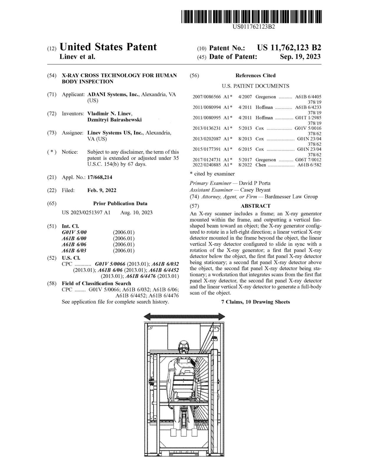
An X-ray scanner includes a frame; an X-ray generator mounted within the frame, and outputting a vertical fan-shaped beam toward an object; the X-ray generator configured to rotate in a left-right direction; a linear vertical X-ray detector mounted in the frame beyond the object, the linear vertical X-ray detector configured to slide in sync with a rotation of the X-ray generator; a first flat panel X-ray detector below the object, the first flat panel X-ray detector being stationary; a second flat panel X-ray detector above the object, the second flat panel X-ray detector being stationary; a workstation that integrates scans from the first flat panel X-ray detector, the second flat panel X-ray detector and the linear vertical X-ray detector to generate a full-body scan of the object.
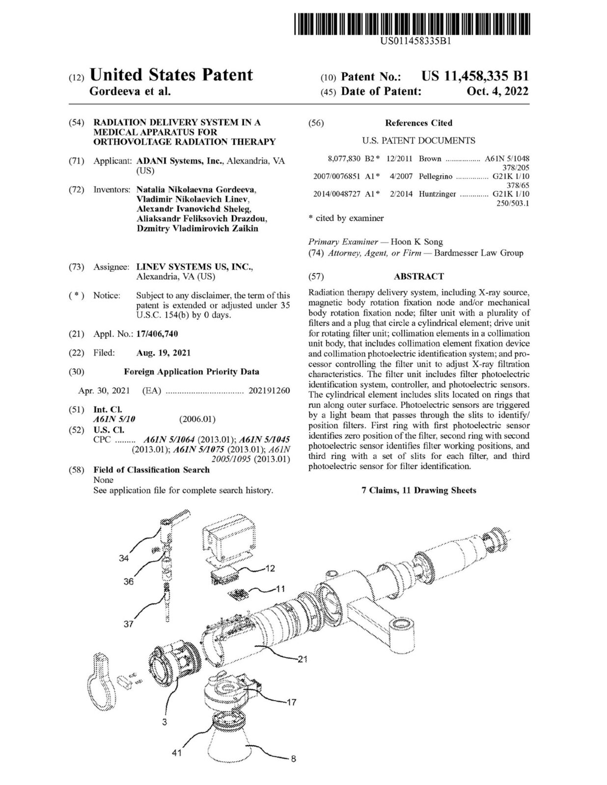
Radiation therapy delivery system, including X-ray source, magnetic body rotation fixation node and/or mechanical body rotation fixation node; filter unit with a plurality of filters and a plug that circle a cylindrical element; drive unit for rotating filter unit; collimation elements in a collimation unit body, that includes collimation element fixation device and collimation photoelectric identification system; and pro- cessor controlling the filter unit to adjust X-ray filtration characteristics. The filter unit includes filter photoelectric identification system, controller, and photoelectric sensors. The cylindrical element includes slits located on rings that run along outer surface. Photoelectric sensors are triggered by a light beam that passes through the slits to identify/ position filters. First ring with first photoelectric sensor identifies zero position of the filter, second ring with second photoelectric sensor identifies filter working positions, and third ring with a set of slits for each filter, and third photoelectric sensor for filter identification.

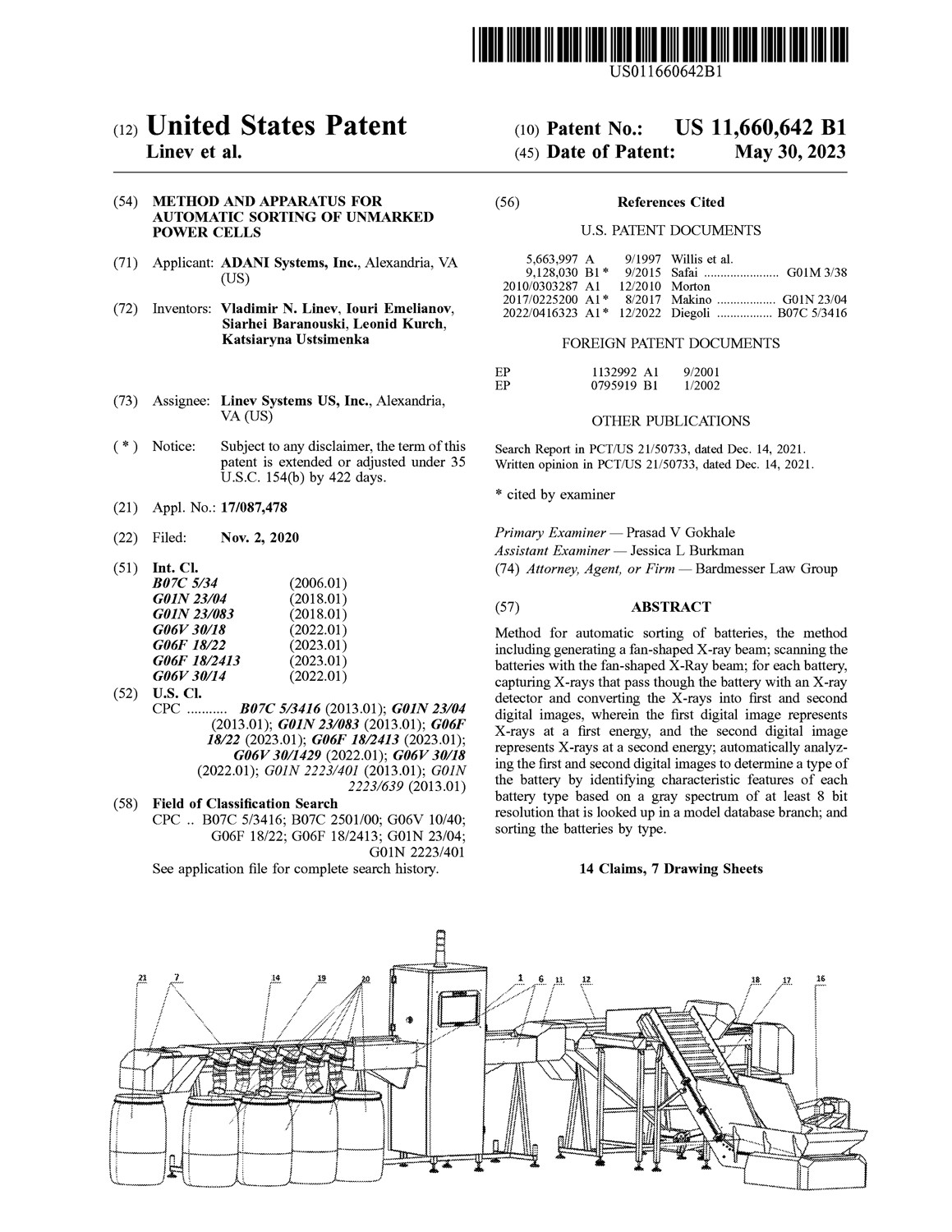
Method for automatic sorting of batteries, the method including generating a fan-shaped X-ray beam; scanning the batteries with the fan-shaped X-Ray beam; for each battery, capturing X-rays that pass though the battery with an X-ray detector and converting the X-rays into first and second digital images, wherein the first digital image represents X-rays at a first energy, and the second digital image represents X-rays at a second energy; automatically analyzing the first and second digital images to determine a type of the battery by identifying characteristic features of each battery type based on a gray spectrum of at least 8 bit resolution that is looked up in a model database branch; and sorting the batteries by type.
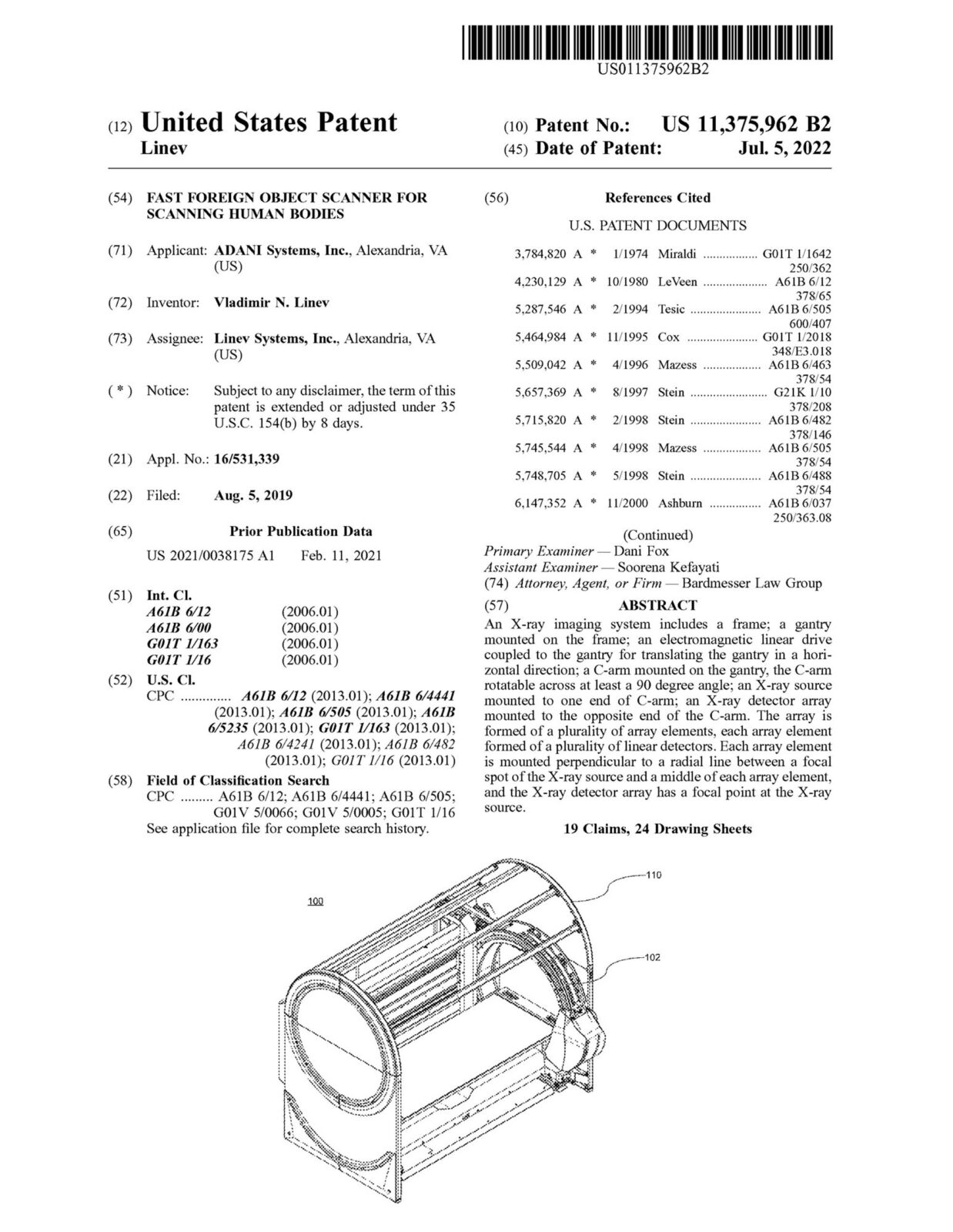
An X-ray imaging system includes a frame; a gantry mounted on the frame; an electromagnetic linear drive coupled to the gantry for translating the gantry in a horizontal direction; a C-arm mounted on the gantry, the C-arm rotatable across at least a 90 degree angle; an X-ray source mounted to one end of C-arm; an X-ray detector array mounted to the opposite end of the C-arm. The array is formed of a plurality of array elements, each array element formed of a plurality of linear detectors. Each array element is mounted perpendicular to a radial line between a focal spot of the X-ray source and a middle of each array element, and the X-ray detector array has a focal point at the X-ray source.

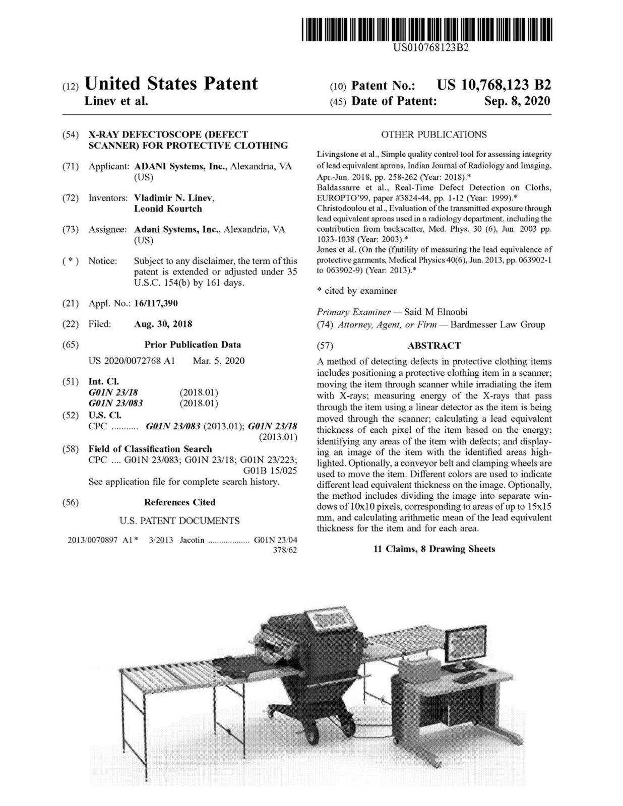
A method of detecting defects in protective clothing items includes positioning a protective clothing item in a scanner; moving the item through scanner while irradiating the item with X-rays; measuring energy of the X-rays that pass through the item using a linear detector as the item is being moved through the scanner; calculating a lead equivalent thickness of each pixel of the item based on the energy; identifying any areas of the item with defects; and displaying an image of the item with the identified areas highlighted. Optionally, a conveyor belt and clamping wheels are used to move the item. Different colors are used to indicate different lead equivalent thickness on the image. Optionally, the method includes dividing the image into separate windows of 10×10 pixels, corresponding to areas of up to 15×15 mm, and calculating arithmetic mean of the lead equivalent thickness for the item and for each area.
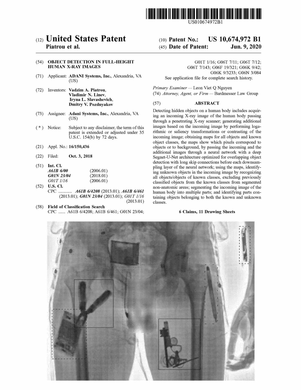
Detecting hidden objects on a human body includes acquiring an incoming X-ray image of the human body passing through a penetrating X-ray scanner; generating additional images based on the incoming image by performing logarithmic or saliency transformations or contrasting of the incoming image; obtaining maps for all objects and known object classes, the maps show which pixels correspond to objects or to background, by passing the incoming and the additional images through a neural network with a deep Segnet-U-Net architecture optimized for overlapping object detection with long skip connections before each downsam- pling layer of the neural network; using the maps, identifying unknown objects in the incoming image by recognizing all objects/objects of known classes, excluding previously classified objects from the known classes from segmented non-anatomic areas; segmenting the incoming image of the human body into multiple parts; and identifying parts con- taining objects belonging to both the known and unknown classes.

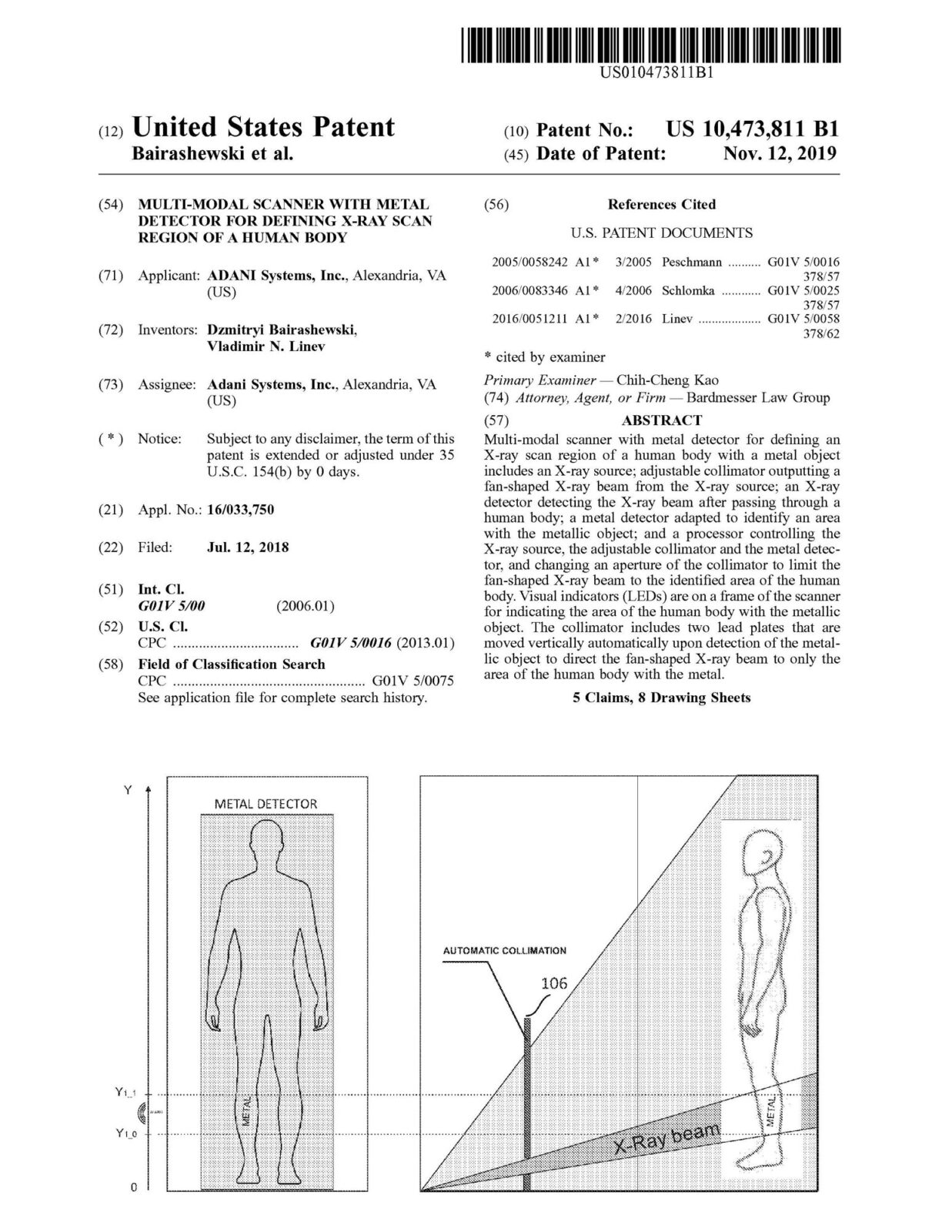
Multi-modal scanner with metal detector for defining an X-ray scan region of a human body with a metal object includes an X-ray source; adjustable collimator outputting a fan-shaped X-ray beam from the X-ray source; an X-ray detector detecting the X-ray beam after passing through a human body; a metal detector adapted to identify an area with the metallic object; and a processor controlling the X-ray source, the adjustable collimator and the metal detector, and changing an aperture of the collimator to limit the fan-shaped X-ray beam to the identified area of the human body. Visual indicators (LEDs) are on a frame of the scanner for indicating the area of the human body with the metallic object. The collimator includes two lead plates that are moved vertically automatically upon detection of the metallic object to direct the fan-shaped X-ray beam to only the area of the human body with the metal.
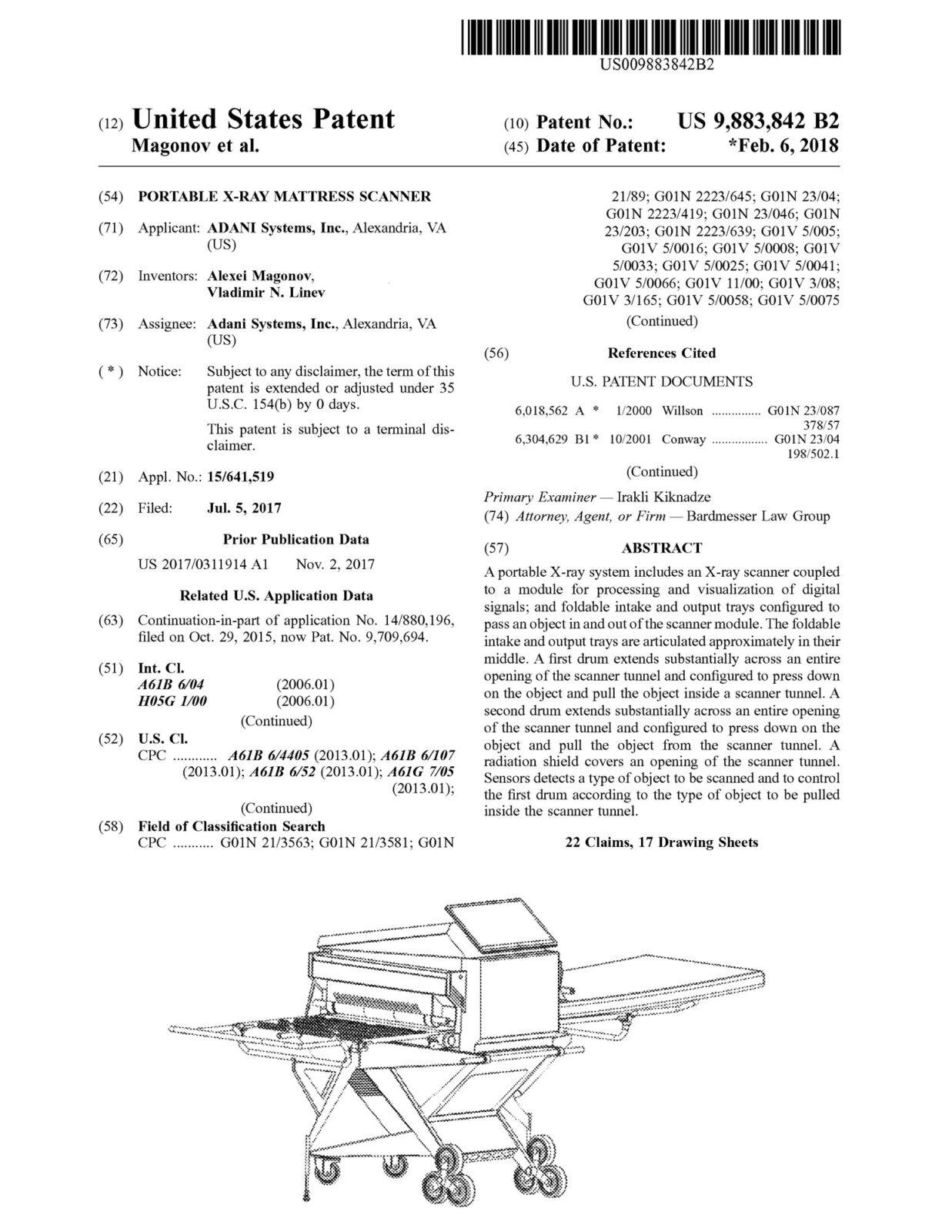
A portable X-ray system includes an X-ray scanner coupled to a module for processing and visualization of digital signals; and foldable intake and output trays configured to pass an object in and out of the scanner module. The foldable intake and output trays are articulated approximately in their middle. A first drum extends substantially across an entire opening of the scanner tunnel and configured to press down on the object and pull the object inside a scanner tunnel. A second drum extends substantially across an entire opening of the scanner tunnel and configured to press down on the object and pull the object from the scanner tunnel. A radiation shield covers an opening of the scanner tunnel. Sensors detects a type of object to be scanned and to control the first drum according to the type of object to be pulled inside the scanner tunnel.

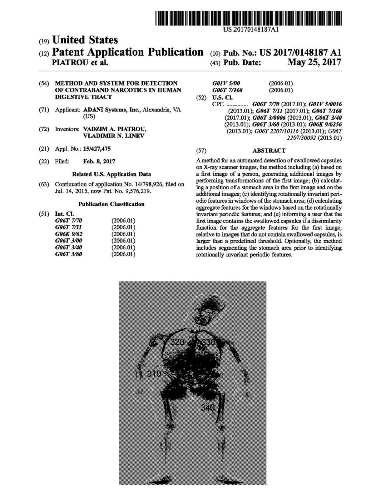
A method for an automated detection of swallowed capsules on X-ray scanner images, the method including (a) based on a first image of a person, generating additional images by performing transformations of the first image; (b) calculating a position of a stomach area in the first image and on the additional images; (c) identifying rotationally invariant periodic features in windows of the stomach area; (d) calculating aggregate features for the windows based on the rotationally invariant periodic features; and (e) informing a user that the first image contains the swallowed capsules if a dissimilarity function for the aggregate features for the first image, relative to images that do not contain swallowed capsules, is larger than a predefined threshold. Optionally, the method includes segmenting the stomach area prior to identifying rotationally invariant periodic features.
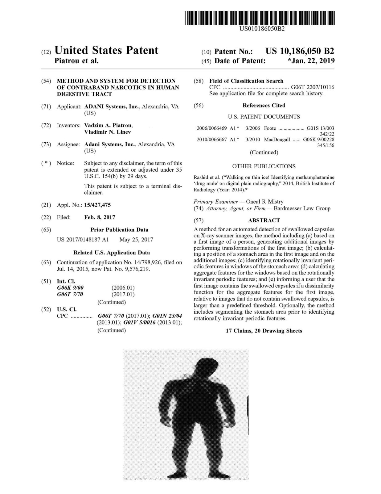
A method for an automated detection of swallowed capsules on X-ray scanner images, the method including (a) based on a first image of a person, generating additional images by performing transformations of the first image; (b) calculat- ing a position of a stomach area in the first image and on the additional images; (c) identifying rotationally invariant peri- odic features in windows of the stomach area; (d) calculating aggregate features for the windows based on the rotationally invariant periodic features; and (e) informing a user that the first image contains the swallowed capsules if a dissimilarity function for the aggregate features for the first image, relative to images that do not contain swallowed capsules, is larger than a predefined threshold. Optionally, the method includes segmenting the stomach area prior to identifying rotationally invariant periodic features.

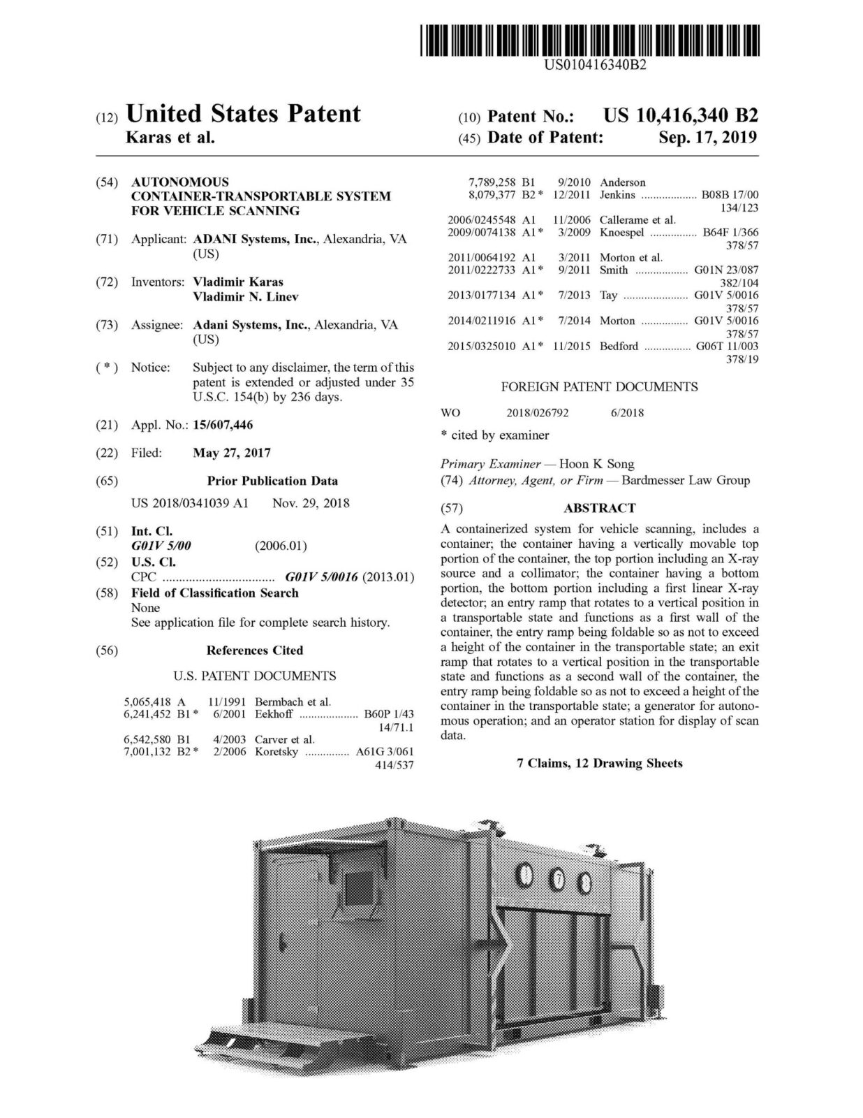
A containerized system for vehicle scanning. includes a container; the container having a vertically movable top portion of the container, the top portion including an X-ray source and a collimator; the container having a bottom portion, the bottom portion including a first linear X-ray detector, an entry ramp that rotates to a vertical position in a transportable state and functions as a first wall of the container, the entry ramp being foldable so as not to exceed a height of the container in the transportable state; an exit ramp that rotates to a vertical position in the transportable state and functions as a second wall of the container, the entry ramp being foldable so as not to exceed a height of the container in the transportable state; a generator for autonomous operation; and an operator station for display of scan data.
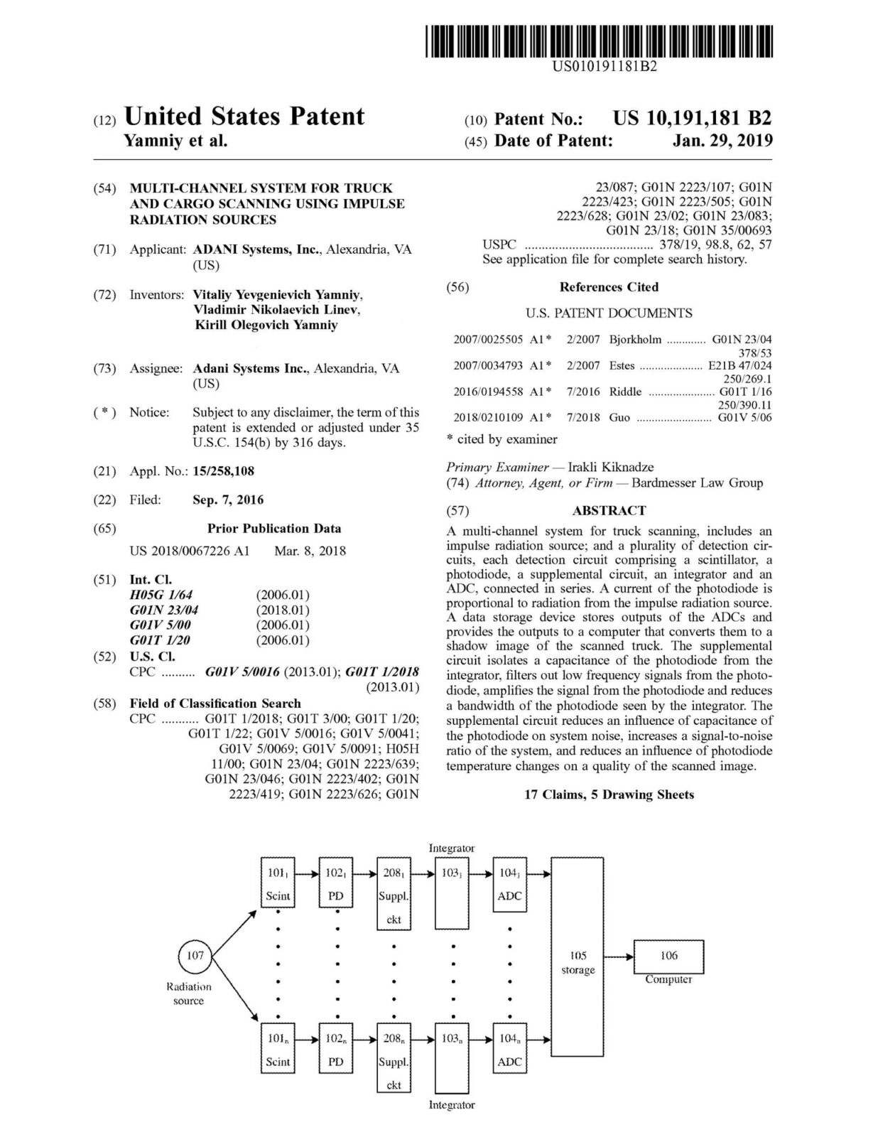
A multi-channel system for truck scanning, includes an impulse radiation source; and a plurality of detection circuits, each detection circuit comprising a scintillator, a photodiode, a supplemental circuit, an integrator and an ADC, connected in series. A current of the photodiode is proportional to radiation from the impulse radiation source. A data storage device stores outputs of the ADCs and provides the outputs to a computer that converts them to a shadow image of the scanned truck. The supplemental circuit isolates a capacitance of the photodiode from the integrator, filters out low frequency signals from the photodiode, amplifies the signal from the photodiode and reduces a bandwidth of the photodiode seen by the integrator. The supplemental circuit reduces an influence of capacitance of the photodiode on system noise, increases a signal-to-noise ratio of the system, and reduces an influence of photodiode temperature changes on a quality of the scanned image.

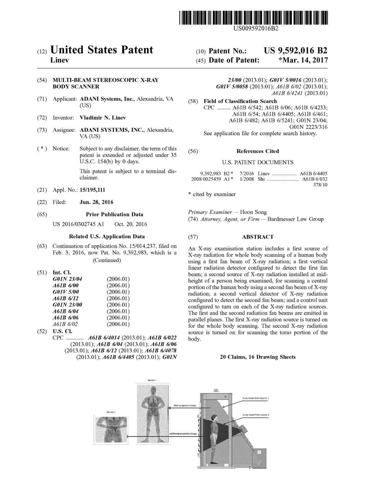
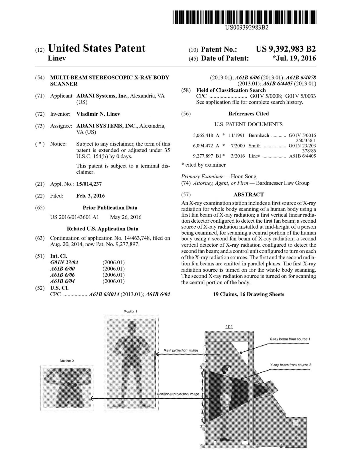
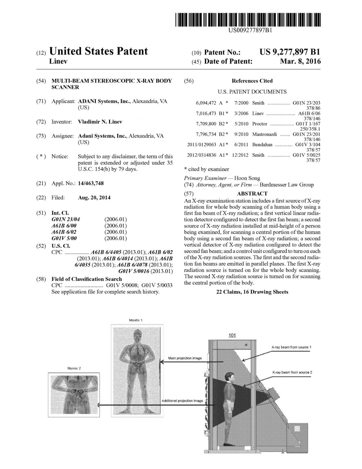
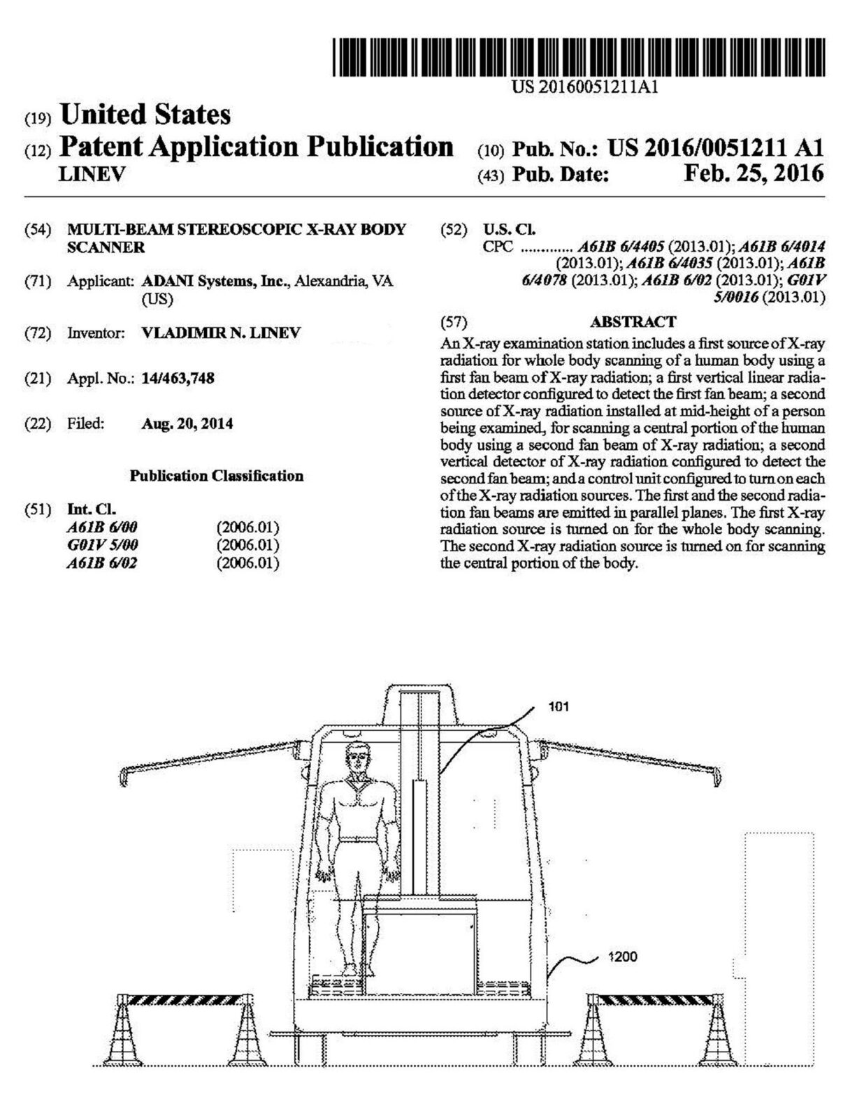
An X-ray examination station includes a first source of X-ray radiation for whole body scanning of a human body using a first fan beam of X-ray radiation; fist vertical linear radiation detector configured to detect the first fan beam: a second source of X-ray radiation installed at mid-height of a person being examined, for scanning a central portion of the human body using a second fan beam of X-ray radiation; a second vertical detector of X-ray radiation configured to detect the second fan beam; and a control unit configured to turn on each of the X-ray radiation sources. The first and the second radiation fan beams are emitted in parallel planes. The first X-ray source is tumed on for the whole body scanning.The second X-ray radiation source is tumed on for scanning the central portion of the body.
An X-ray examination station includes a first source of X-ray radiation for whole body scanning of a human body using a first fan beam of X-ray radiation; fist vertical linear radiation detector configured to detect the first fan beam: a second source of X-ray radiation installed at mid-height of a person being examined, for scanning a central portion of the human body using a second fan beam of X-ray radiation; a second vertical detector of X-ray radiation configured to detect the second fan beam; and a control unit configured to turn on each of the X-ray radiation sources. The first and the second radiation fan beams are emitted in parallel planes. The first X-ray source is tumed on for the whole body scanning.The second X-ray radiation source is tumed on for scanning the central portion of the body.

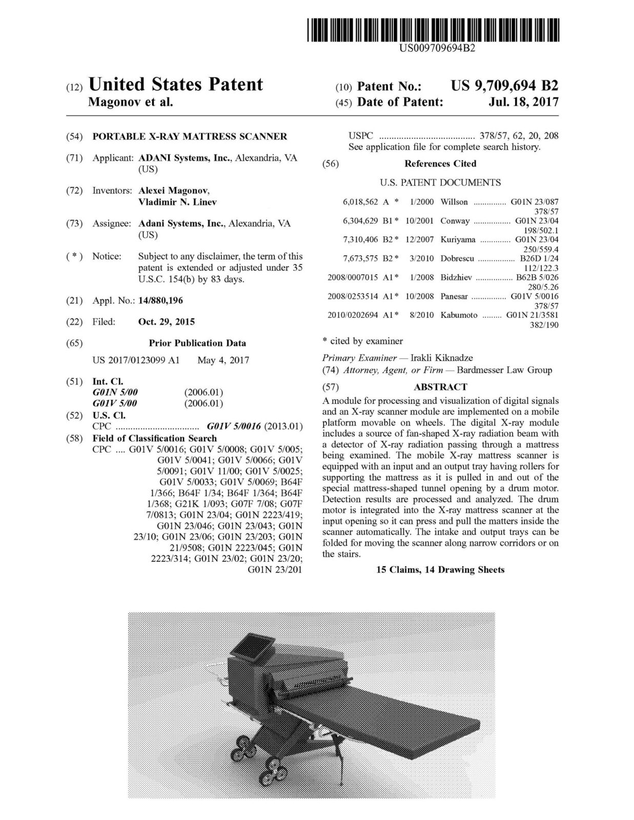
A module for processing and visualization of digital signals and an X-ray scanner module are implemented on a mobile platform movable on wheels. The digital X-ray module includes a source of fan-shaped X-ray radiation beam with a detector of X-ray radiation passing through a mattress being examined. The mobile X-ray mattress scanner is equipped with an input and an output tray having rollers for supporting the mattress as it is pulled in and out of the special mattress-shaped tunnel opening by a drum motor. Detection results are processed and analyzed. The drum motor is integrated into the X-ray mattress scanner at the input opening so it can press and pull the matters inside the scanner automatically. The intake and output trays can be folded for moving the scanner along narrow corridors or on the stairs.
A method for automated detection of illegal substances smuggled inside internal cavities of a passenger, e.g., as capsules. The method provides for an automated detection of narcotics hidden in a passenger’s stomach area using pictures produced by an X-ray scanner. According to an exemplary embodiment, throughput of the scanner is increased by an automated detection algorithm, which takes less time than visual analysis by an operator. The operator is only involved in cases when narcotics are detected. The automated detection method has a consistent precision, because the effects of tiredness of the operator are eliminated. Efficiency and costs of the process are improved, since fewer qualified operators can service several scanners.

An X-ray examination station includes a first source of X-ray radiation for whole body scanning of a human body using a first fan beam of X-ray radiation; fist vertical linear radiation detector configured to detect the first fan beam: a second source of X-ray radiation installed at mid-height of a person being examined, for scanning a central portion of the human body using a second fan beam of X-ray radiation; a second vertical detector of X-ray radiation configured to detect the second fan beam; and a control unit configured to turn on each of the X-ray radiation sources. The first and the second radiation fan beams are emitted in parallel planes. The first X-ray source is tumed on for the whole body scanning.The second X-ray radiation source is tumed on for scanning the central portion of the body.
An X-ray examination station includes a first source of X-ray radiation for whole body scanning of a human body using a first fan beam of X-ray radiation; a first vertical linear radiation detector configured to detect the first fan beam; a second source of X-ray radiation installed at mid-height of a person being examined, for scanning a central portion of the human body using a second fan beam of X-ray radiation; a second vertical detector of X-ray radiation configured to detect the second fan beam; and a control unit configured to turn on each of the X-ray radiation sources. The first and the second radiation fan beams are emitted in parallel planes. The first X-ray radiation source is turned on for the whole body scanning. The second X-ray radiation source is turned on for scanning the central portion of the body.

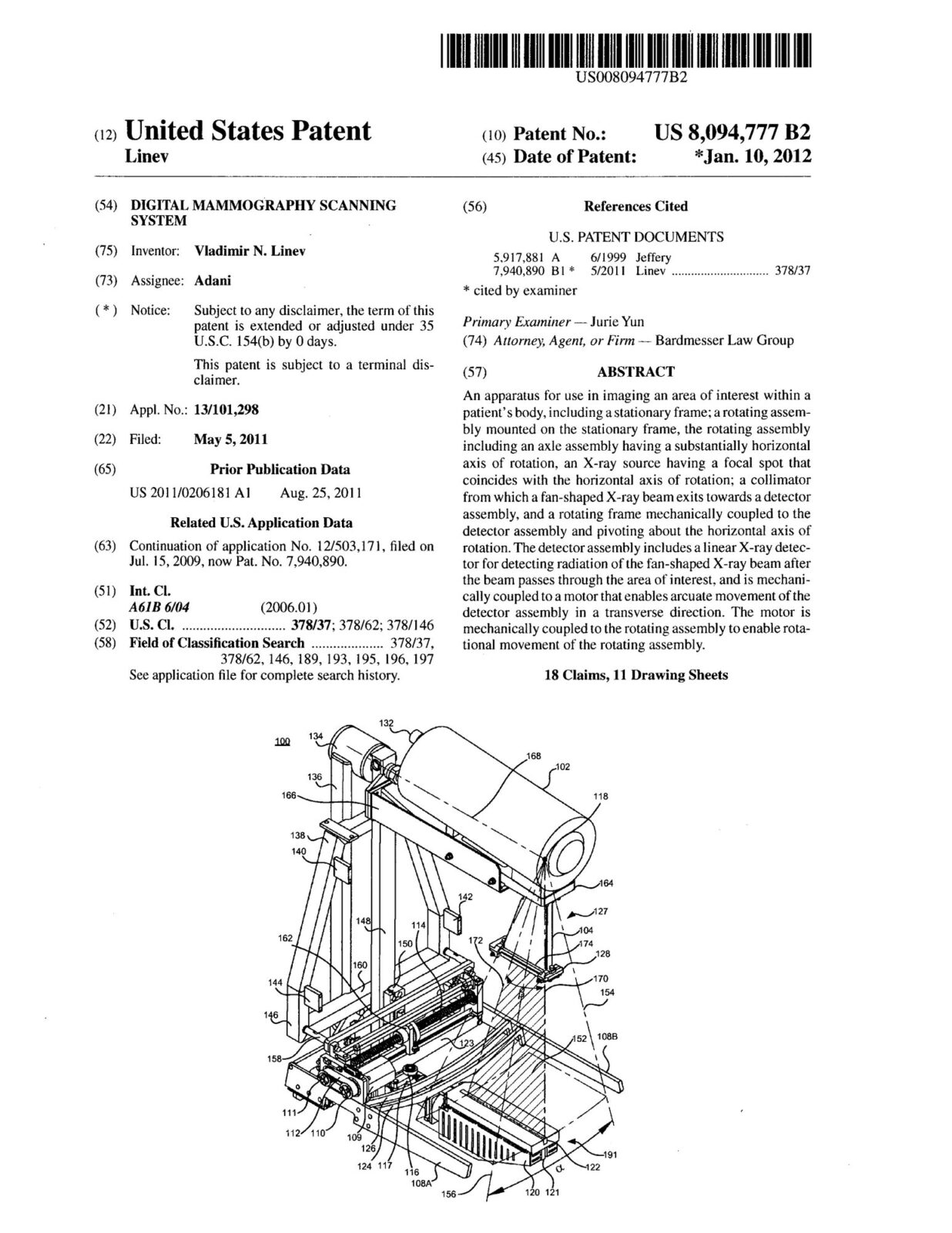
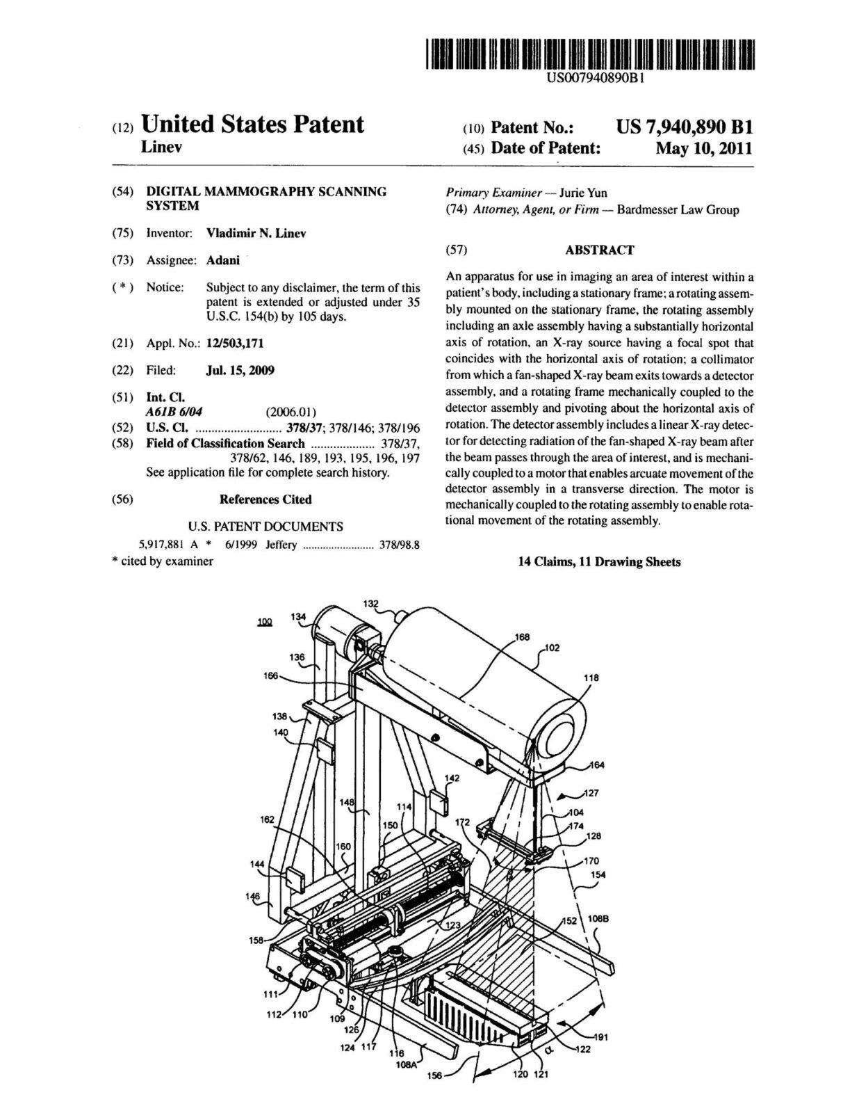
An apparatus for use in imaging an area of interest within a patient’s body, including a stationary frame: a rotating assembly mounted on the stationary frame, the rotating assembly including an axle assembly having a substantially horizontal axis of rotation, an X-ray source having a focal spot that coincides with the horizontal axis of rotation: a collimator from which a fan-shaped X-ray beam exits towards a detector assembly, and a rotating frame mechanically coupled to the detector assembly and pivoting about the horizontal axis of rotation. The detector assembly includes a linear X-ray detector for detecting radiation of the fan-shaped X-ray beam after the beam passes through the area of interest, and is mechanically coupled to a motor that enables arcuate movement of the detector assembly in a transverse direction. The motor is mechanically coupled to the rotating assembly to enable rotational movement of the rotating assembly.
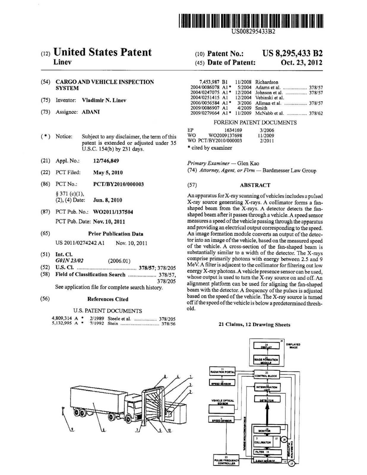
An apparatus for X-ray scanning of vehicles includes a pulsed X-ray source generating X-rays. A collimator forms a fan-shaped beam from the X-rays. A detector detects the fan-shaped beam after it passes through a vehicle. A speed sensor measures a speed of the vehicle passing through the apparatus and providing an electrical output corresponding to the speed. An image formation module converts an output of the detector into an image of the vehicle, based on the measured speed of the vehicle. A cross-section of the fan-shaped beam is substantially similar to a width of the detector. The X-rays comprise primarily photons with energy between 2.5 and 9 MeV. A filter is adjacent to the collimator for filtering out low energy X-ray photons. A vehicle presence sensor can be used, whose output is used to turn the X-ray source on and off. An alignment platform can be used for aligning the fan-shaped beam with the detector. A frequency of the pulses is adjusted based on the speed of the vehicle. The X-ray source is turned off if the speed of the vehicle is below a predetermined threshold.

An apparatus for use in imaging an area of interest within a patient’s body, including a stationary frame: a rotating assembly mounted on the stationary frame, the rotating assembly including an axle assembly having a substantially horizontal axis of rotation, an X-ray source having a focal spot that coincides with the horizontal axis of rotation: a collimator from which a fan-shaped X-ray beam exits towards a detector assembly, and a rotating frame mechanically coupled to the detector assembly and pivoting about the horizontal axis of rotation. The detector assembly includes a linear X-ray detector for detecting radiation of the fan-shaped X-ray beam after the beam passes through the area of interest, and is mechanically coupled to a motor that enables arcuate movement of the detector assembly in a transverse direction. The motor is mechanically coupled to the rotating assembly to enable rotational movement of the rotating assembly.
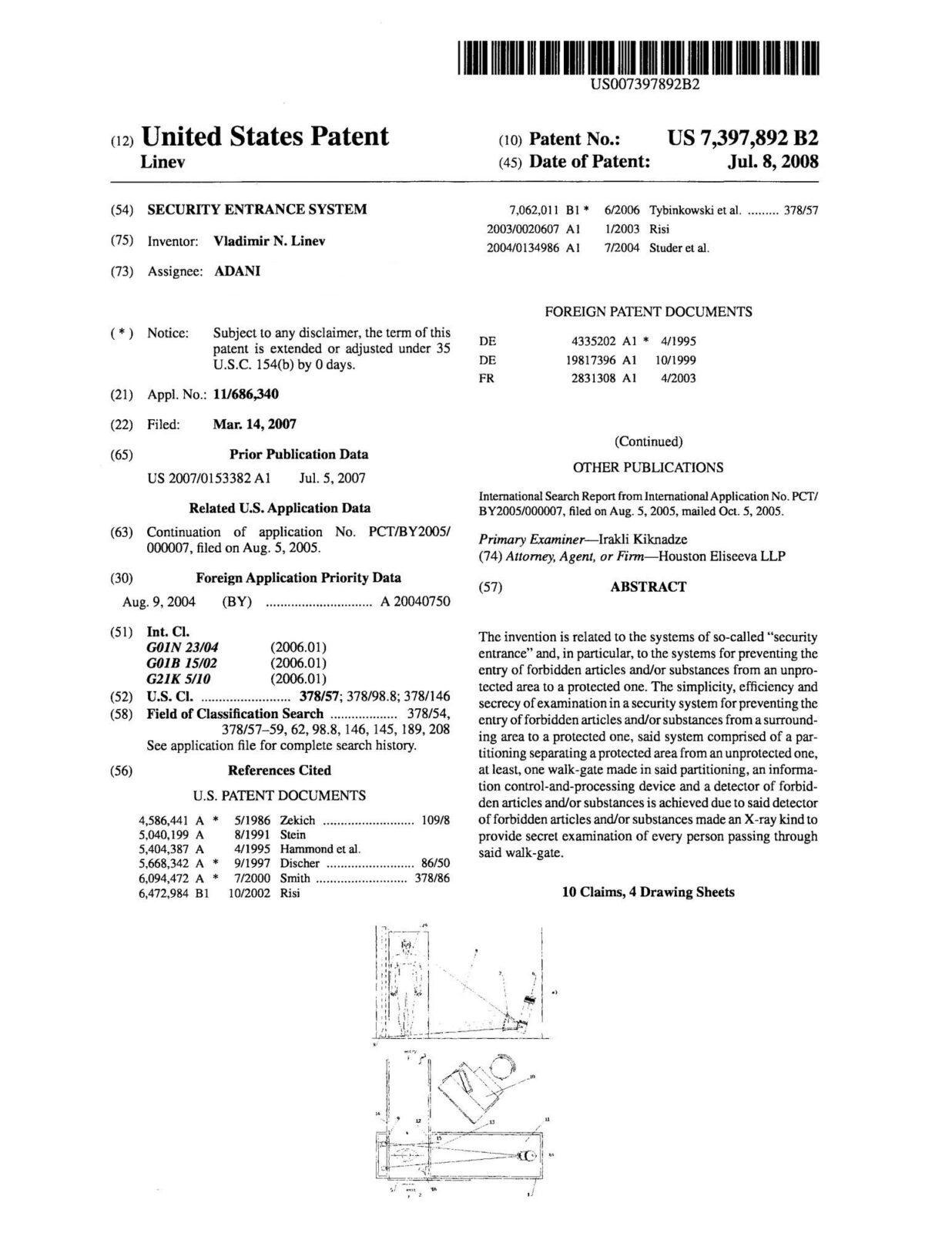
The invention is related to the systems of so-called “security entrance” and, in particular, to the systems for preventing the entry of forbidden articles and/or substances from an unprotected area to a protected one. The simplicity, efficiency and secrecy of examination in a security system for preventing the entry of forbidden articles and/or substances from a surrounding area to a protected one, said system comprised of a par titioning separating a protected area from an unprotected one, at least, one walk-gate made in said partitioning, an information control-and-processing device and a detector of forbidden articles and/or substances is achieved due to said detector of forbidden articles and/or substances made an X-ray kind to provide secret examination of every person passing through said walk-gate.

The ornamental design for an x-ray apparatus for body scanning, as shown and described. An apparatus for x-ray scanning a body, such as that of a human patient.
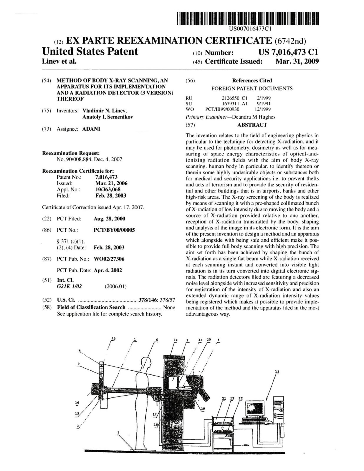
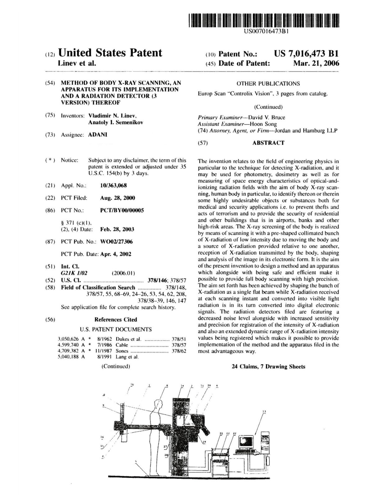
The invention relates to the field of engineering physics in particular to the technique for detecting X-radiation, and it may be used for photometry, dosimetry as well as for measuring of space energy characteristics of optical-and-ionizing radiation fields with the aim of body X-ray scanning, human body in particular, to identify thereon or therein some highly undesirable objects or substances both for medical and security applications i.e. to prevent thefts and acts of terrorism and to provide the security of residential and other buildings that is in airports, banks and other high-risk areas. The X-ray screening of the body is realized by means of scanning it with a pre-shaped collimated bunch of X-radiation of low intensity due to moving the body and a source of X-radiation provided relative to one another, reception of X-radiation transmitted by the body, shaping and analysis of the image in its electronic form.

The invention relates to the field of engineering physics in particular to the technique for detecting X-radiation, and it may be used for photometry, dosimetry as well as for measuring of space energy characteristics of optical-and-ionizing radiation fields with the aim of body X-ray scanning, human body in particular, to identify thereon or therein some highly undesirable objects or substances both for medical and security applications i.e. to prevent thefts and acts of terrorism and to provide the security of residential and other buildings that is in airports, banks and other high-risk areas. The X-ray screening of the body is realized by means of scanning it with a pre-shaped collimated bunch of X-radiation of low intensity due to moving the body and a source of X-radiation provided relative to one another, reception of X-radiation transmitted by the body, shaping and analysis of the image in its electronic form.


